Chest Muscle Anatomy Diagram : Cat muscles - Human Anatomy And Physiology with Alvarez at ... : Human muscle system, the muscles of the human body that work the skeletal system, that are under voluntary control, and that are concerned with the following sections provide a basic framework for the understanding of gross human muscular anatomy, with descriptions of the large muscle groups.
Chest Muscle Anatomy Diagram : Cat muscles - Human Anatomy And Physiology with Alvarez at ... : Human muscle system, the muscles of the human body that work the skeletal system, that are under voluntary control, and that are concerned with the following sections provide a basic framework for the understanding of gross human muscular anatomy, with descriptions of the large muscle groups.. Muscle anatomy types of movement all muscles exert their force by pulling between at least two points of attachment. Anatomical diagram showing a front view of muscles in the human body. There are over 630 muscles in the human body; Want to learn more about it? Muscles that act on the chest.
Muscles that act on the chest. Anatomy • free medical books. Human anatomy diagram shoulder anatomy shoulder muscles shoulder muscles and chest. It forms the bulk of the chest area and can be easily. The movement that results from contraction is called the action of the muscle.

Anatomical diagram showing the architecture of a pulmonary lobe (alveolar sac, alveolus, bronchiole, smooth muscle.)
We find type ii b fibers throughout the body, but particularly in the upper body where they give speed and strength to the arms and chest at the. Anatomical diagram showing a front view of muscles in the human body. The pectoralis minor muscle (not shown in the diagram) is located underneath the pectoralis major muscle, attaching to the coracoid. It forms the bulk of the chest area and can be easily. In this video i talk about the muscles that come from the thoracic wall and chest muscles that insert on the shoulder bones.✅. Learn vocabulary, terms and more with flashcards, games and other study tools. Learn to draw the knee, feet and toes following this detailed and thorough online free art book. The serratus anterior is located more laterally in the chest wall and forms the medial border of the axilla region. Choose from over a million free vectors, clipart graphics, vector art images, design templates, and illustrations created by artists worldwide! The soleus connects your lower leg bones to your heel, but it also gives your heart some help by pumping blood back. To get started, choose a muscle group either on the muscle chart. A massive chest anchors the upper body and enhances the. Find out more about the individual muscles within the chest anatomy by clicking their respective links throughout this page.
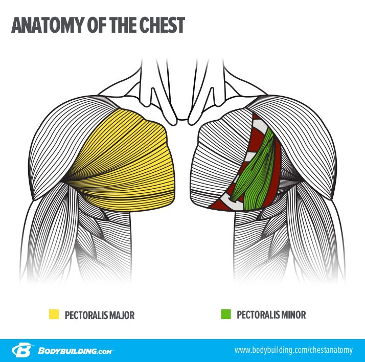
Almost all muscles cross at least one joint (moveable connection between two bones) and cause an action across that joint. The chest anatomy includes the pectoralis major, pectoralis minor and the serratus anterior. Chest muscle anatomy and exercises, chest muscle chart, chest muscle diagram workout, muscle diagram of chest, muscle diagram muscle anatomy labeled 12 photos of the muscle anatomy labeled muscle anatomy diagrams, muscle anatomy labeling exercises, muscle anatomy. Meet your pectoralis major and pectoralis minor. Muscles that act on the chest.
Typically, one attachment remains stationary and is called the origin and the other attachment moves.
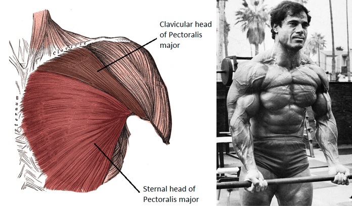
We think this is the most useful anatomy picture that. To get started, choose a muscle group either on the muscle chart. Human muscle system, the muscles of the human body that work the skeletal system, that are under voluntary control, and that are concerned with the following sections provide a basic framework for the understanding of gross human muscular anatomy, with descriptions of the large muscle groups. The pectoralis minor muscle (not shown in the diagram) is located underneath the pectoralis major muscle, attaching to the coracoid. The pectoralis major muscles (also known as the pecs) are located on the front of the rib cage, and form the major muscles of the chest. Anatomical diagram showing a front view of muscles in the human body. The muscle consists of several strips, which originate from the lateral aspects of. Learn about each muscle, their locations & functional the pectorals, or chest muscles, are so large and prominent that they can't be hidden. Chest muscles anatomy for bodybuilders. This page provides an overview of the chest muscle group. Chest muscle anatomy and exercises, chest muscle chart, chest muscle diagram workout, muscle diagram of chest, muscle diagram muscle anatomy labeled 12 photos of the muscle anatomy labeled muscle anatomy diagrams, muscle anatomy labeling exercises, muscle anatomy. Start studying chest muscles anatomy. Learn to draw the knee, feet and toes following this detailed and thorough online free art book.
Want to learn more about it? Note how the basilar segmental bronchi are oriented from lateral to medial. This quiz focuses on the 23 largest muscles—the ones that account for most of your mobility and strength. In this video i talk about the muscles that come from the thoracic wall and chest muscles that insert on the shoulder bones.✅. Diagrams photos diagram of the chest human anatomy.
Meet your pectoralis major and pectoralis minor.
The two sides connect at the sternum, or breastbone. Groin muscles diagram anterior muscles diagram muscle diagram anterior muscular system. Constructive anatomy by canadian artist george bridgman, free art book to read online. They are categorized by the muscles which they affect (primary and secondary), as well as the equipment required. Typically, one attachment remains stationary and is called the origin and the other attachment moves. The movement that results from contraction is called the action of the muscle. Muscle anatomy types of movement all muscles exert their force by pulling between at least two points of attachment. Download human muscle anatomy diagram vector art. The muscle consists of several strips, which originate from the lateral aspects of. Note how the basilar segmental bronchi are oriented from lateral to medial. The soleus connects your lower leg bones to your heel, but it also gives your heart some help by pumping blood back. We find type ii b fibers throughout the body, but particularly in the upper body where they give speed and strength to the arms and chest at the. Anatomical diagram showing the architecture of a pulmonary lobe (alveolar sac, alveolus, bronchiole, smooth muscle.)


Komentar
Posting Komentar